The modern cell theory, one of the fundamental generalizations of biology, holds that:
- All organisms are composed of one or more cells.
- New cells come from pre‐existing cells; lifeforms today have descended in unbroken continuity from the first primitive cells that arose on earth more than 3.5 billion years ago.
- Hereditary information passes from parent cell to daughter cell.
- The fundamental biochemical reactions of life take place within cells.
Microscopes. Cytology—the scientific study of cells—has progressed simultaneously with the development of better, more powerful microscopes and cell preparation techniques. Light microscopes, invented in the 1500s, today can magnify in the range of 100x to 1000x and are at the maximum of their resolving power at about 0.2 Φm (micrometer = 1/ 1,000,000 meter). They are limited from further improvement by the wavelength of visible light that is used to create the image (the shorter the wavelength, the greater the resolution).
Beams of electrons have shorter wavelengths than visible light, hence electron microscopes (which focus streams of electrons) are able to achieve resolutions of 2–0.4 nm (nanometer = 1/ 1,000,000,000 meter = 1/1000 Φm) and magnifications of over 100,000x. One type of electron microscope, the transmission electron microscope ( TEM), focuses a beam of electrons through an object much as a light beam is used in light microscopes, whereas a scanning electron microscope ( SEM) forms an image from electrons that are scattered from the surface of a specimen.
Laboratory techniques. In light microscopy dyes that stain particular chemicals are commonly used to identify cellular structures in thin sections of tissue mounted on microscope slides. Specimens observed by electron microscopy are cut in much thinner sections (electrons don't penetrate material well) and examined in a vacuum. These preparations cause problems of interpretation of the microscopic image. Does the material really look like this in living cells or has preparation altered its appearance? Living material can not be examined using electron microscopes so the answer is moot.
Today's understanding of cells is based on two approaches: 1.) the sometimes very difficult biochemical analyses of cells and 2.) the increasing greater magnification of their constituent parts.
Plant cells vary in size from about 7 Φm—the diameter of dividing cells at the tips of roots and shoots—to fiber cells a few micrometers wide but a meter or more in length. Most plant (and animal) cells are between 10–100 Φm and thus too small to be seen without magnification since human eyes have a resolving power (resolution) of about 100 Φm. (Resolving power is the ability to see adjacent objects as separate; magnification simply makes things larger.
Two things closer together than 100 Φm will look like one to the human eye unless a microscope with a resolving power of less than 100 Φm is used.
Plant cells are separated from their environment by a cell wall inside of which a semi‐liquid plasma membrane surrounds the cytoplasm and the nucleus. As cells grow, their volume increases faster than their surface area, a geometric reality (volume increases as the cube of the diameter, surface as the square of the diameter in the surface:volume ratio). Since materials used and released in the metabolism of the cell must pass through its surface, a functional upper size limit to growth is reached; cells can grow no larger in volume than the membrane area can support. Distribute the same volume of cytoplasm contained in one large cell into several smaller cells and the surface area increases, the cell can function, and ultimately, multicellular organisms result. Multicellular bodies have advantages over unicellular ones, e.g. cells can specialize to perform specific biochemical functions, different kinds of structural materials and structures can be produced, and the control center of the cell, the nucleus, is more efficient with fewer activities to control.
All cells have
- An outer membrane, the plasma membrane (also called the cell membrane or plasmalemma), composed of a lipid bilayer in which proteins are embedded.
- Genetic material, found in molecules of deoxyribonucleic acid (DNA).
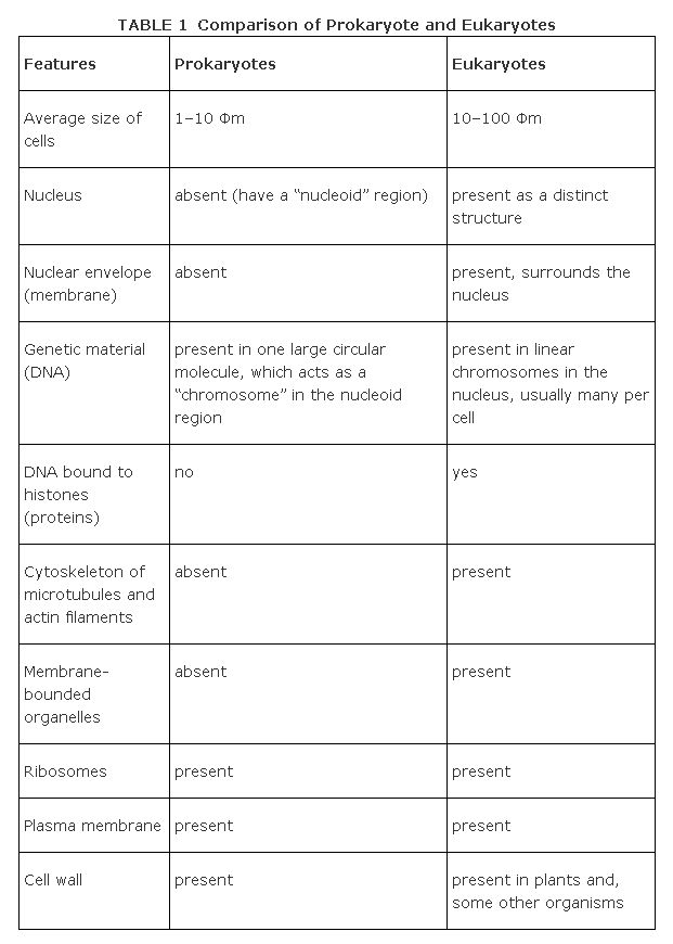
The fundamental separation. Based on cellular structure, the living world can be divided into two kinds of organisms: the prokaryotes and the eukaryotes. The cells of prokaryotes (the bacteria) lack a nucleus and other membrane-bounded cellorganelles whereas those of the eukaryotes—the rest of the living world—have nuclei and a complex internal structure with membrane-bounded cell organelles. Table 1compares features of the cells of the two basic types.
Origin of the eukaryote type. Biologists in general agree that early organisms were prokaryotes that developed ways to survive on a young Earth whose atmosphere and conditions were much different from today's. Descendants of these ancient prokaryotes—the organisms of today—carry in their genes solutions to old problems as well as modifications made over 3.5 billion years in response to changing Earth conditions.
The mitochondria and chloroplasts of plants differ from other organelles in their semi-independence. In many ways they resemble bacteria: Both bacteria and the organellescan divide, their DNA is organized similarly, and both have small ribosomes. Additionally, antibiotics inhibit protein synthesis in ribosomes of mitochondria, chloroplasts, and bacteria, but have no effect on synthesis in cytosomal ribosomes of eukaryotic cells. These structural and physiological differences lead some biologists to postulate the serial endosymbiotic theory of organelles in eukaryote cells. (Asymbiosis is the close association and living together of individuals of different species.) The theory suggests that, early in the development of life on Earth, what are now organelles of larger, eukaryotic cells were simple, free-living prokaryotes. At some point in time, each independently developed a symbiotic relationship with larger, heterotrophic cells bringing to the partnership energy-capturing and -converting abilities and receiving in trade a physical home that provided the chemical necessities for life and protection from severe environmental stresses. The combination proved successful. Cells with the combined attributes were able to colonize previously hostile environments and the stage was set for the invasion of land and development of green, multicellular, terrestrial plants. The process occurred repeatedly—it was a serial endosymbiosis.